Gel electrophoresis is a method that separates (based on size, electrical charge and other physical properties) macromolecules such as nucleic acids or proteins.
The electrophoresis term is used to describe the migration of charged particle under the influence of an electric field. Thus gel electrophoresis refers is the technique in which molecules are forced across a span of gel, motivated by an electrical current. On either end of the gel there are activated electrodes that provide the driving force. Therefore a molecule's properties especially the possession of ionisable groups, determine how rapidly an electric field can move the molecule through a gelatinous medium.
One very important application for gel electrophoresis is in DNA Technology. Biotechnology has for thousands of years been used by people who have used yeast to make flour into bread and grapes into wine. We are now using biotechnology to study the basic processes of life, diagnose illnesses, and develop new treatments for diseases. Some of the tools of biotechnology are natural components of cells. For example restriction enzymes are made by bacteria to protect themselves from viruses. They inactivate the viral DNA by cutting it in specific places. DNA ligase is an enzyme that exists in all cells and is responsible for joining together strands of DNA. Restriction enzymes can be used to cut DNA at specific sequences called recognition sites. They then rejoin the cut strands with DNA ligase to new combinations of genes. Recombinant DNA sequences contain genes from two or more organisms.
This technique has allowed researchers to gain the ability to diagnose diseases such as sickle cell anemia, cystic fibrosis, and Huntington's chorea early in the course of the disease. Many researchers are also applying the techniques of biotechnology to find new treatments for genetic diseases.
DNA technology has triggered research advances in almost all fields of biology. Currently hundreds of useful products are produced by genetic engineering. It has become routine to combine genes from different sources, usually different species--in test tubes, and then transfer this recombinant DNA into living cells where it can be replicated and expressed.
The most important achievements resulting from recombinant DNA technology have been advances in our basic understanding of eukaryotic molecular biology. For example, only through the use of gene-splicing techniques have the details of eukaryotics gene arrangement and regulation been opened to experimental analysis.
Gel Electrophoresis is one of the staple tools in molecular biology and is of critical value in many aspects of genetic manipulation and study. One use is the identification of particular DNA molecules by the band patterns they yield in gel electrophoresis after being cut with various restriction enzymes. Viral DNA, plasmid DNA, and particular segments of chromosomal DNA can all be identified in this way. Another use is the isolation and purification of individual fragments containing interesting genes, which can be recovered from the gel with full biological activity.
Gel electrophoresis makes it possible to determine the genetic difference and the evolutionary relationship among species of plants and animals. Using this technology it is possible to separate and identify protein molecules that differ by as little as a single amino acid.
Paid To Click


Custom Search
Wednesday, January 19, 2011
Tuesday, January 18, 2011
Free Ebook Download Fundamentals of Biochemical Engineering

The biology, biotechnology, chemistry, pharmacy and chemical engineering students at various universtiy and engineering institutions are required to take the Biochemical Engineering course either as an elective or compulsory subject. This book is written keeping in mind the need for a text book on afore subject for students from both engineering and biology backgrounds. The main feature of this book is that it contains the solved problems, which help the students to understand the subject better. The book is divided into three sections: Enzyme mediated bioprocess, whole cell mediated bioprocess and the engineering principle in bioprocess.
DOWNLOAD HERE
DOWNLOAD HERE
Free Ebook Download Immunobiology 6th Edition

Charles A. Janeway, Paul Travers, Mark Walport and Mark Shlomchik, "Immunobiology"
Publisher: Garland Science | 2004 | ISBN: 0815341016 | File type: PDF | 910 pages | 18.4 mb
Immunobiology, Sixth Edition guides the reader through the immune system in all its aspects - from the first engagement of innate immunity to the generation of the adaptive immune response and its clinical consequences. The Sixth Edition has been thoroughly revised and updated, and now includes end-of-chapter questions. Immunobiology sets the standard for currency and authority with its clear writing style and organization, full-color art program, scientific accuracy, frequent updates, and consistent viewpoint - that of the host's interaction with an environment containing many species of potentially harmful microorganisms.
The Sixth Edition of Immunobiology includes a CD-ROM with original immunological animations based on figures in the book and videos selected from visually compelling experiments. All the animations and videos are accompanied by a voice-over narration. The CD also contains an archive of all the figures in the book, loaded into PowerPoint presentations. There is one presentation for each chapter of the book, and figures follow the order of the chapter.
DOWNLOAD HERE
Thursday, January 13, 2011
Combination of Gene Therapy and Chemotherapy Stops Kidney Cancer in Mouse Model
A novel therapeutic approach combining a modified viral vector and a small molecular weight drug produced promising results in a mouse model of human kidney cancer.
Investigators at the Virginia Commonwealth University (Richmond, USA) created a unique adenovirus vector by combining the tail and shaft domains of a serotype 5 virus and the knob domain of a serotype 3 virus. This Ad.5/3 adenovirus was then loaded with the gene needed to express the cancer-killing protein MDA-7/IL-24.
The viral vector was administered to mice bearing human renal carcinoma cells (RCCs), alone or together with the drug sorafenib, a small molecular weight inhibitor of several tyrosine protein kinases. Sorafenib, which is unique in targeting the Raf/Mek/Erk pathway (MAP Kinase pathway), has already been approved by the [US] Food and Drug Administration (FDA) for the treatment of renal carcinoma.
Results published in the December 15, 2010, issue of the journal Cancer Biology & Therapy revealed that infection with the Ad.5/3-mda-7 vector caused kidney cancer cells and normal cells lining the kidneys to secrete MDA-7/IL-24. MDA-7/IL-24 quickly stopped the growth of the primary tumor. As the infected cells continued to secrete MDA-7/IL-24, it entered the blood stream and eventually stopped the growth of a second, distinct tumor not directly infected by the adenovirus. Only renal carcinoma cells were destroyed by this “toxic bystander effect”; normal cells were unaffected. Sorafenib enhanced MDA-7/IL-24 toxicity and significantly increased its antitumor effects in the mouse model.
“While further research is needed, this therapy could be a novel and effective way to treat metastatic kidney cancer and prolong patient survival,” said senior author Dr. Paul Dent, professor of biochemistry at Virginia Commonwealth University. “This is the first study to clearly define that gene therapeutic delivery of MDA-7/IL-24 in kidney cancer should be explored in the clinic, especially since we have demonstrated an established, FDA-approved drug enhances its toxicity to cancer cells.”
Wednesday, January 12, 2011
Tuesday, January 11, 2011
SDS-PAGE Preparation
The purpose of SDS-PAGE is to separate proteins according to their size, and no other physical feature. In order to understand how this works, we have to understand the two halves of the name: SDS and PAGE.
SDS
Since we are trying to separate many different protein molecules of different shapes and sizes, we first want to denatured so that the proteins no longer have any secondary, tertiary or quaternary structure (i.e. we want them to retain only their primary amino acid structure). Consider two proteins that are each 500 amino acids long but one is shaped like a closed umbrella whle the other one looks like an open umbrella. If you tried to run down the street with both of these molecules under your arms, which one would be more likely to slow you down, even though they weigh exactly the same? This analogy illustrates mass and the 3D structure of a molecule will detrmine how well it can move through an environment. We use SDS to denature all proteins to the same linear shape.
Since we are trying to separate many different protein molecules of different shapes and sizes, we first want to denatured so that the proteins no longer have any secondary, tertiary or quaternary structure (i.e. we want them to retain only their primary amino acid structure). Consider two proteins that are each 500 amino acids long but one is shaped like a closed umbrella whle the other one looks like an open umbrella. If you tried to run down the street with both of these molecules under your arms, which one would be more likely to slow you down, even though they weigh exactly the same? This analogy illustrates mass and the 3D structure of a molecule will detrmine how well it can move through an environment. We use SDS to denature all proteins to the same linear shape.
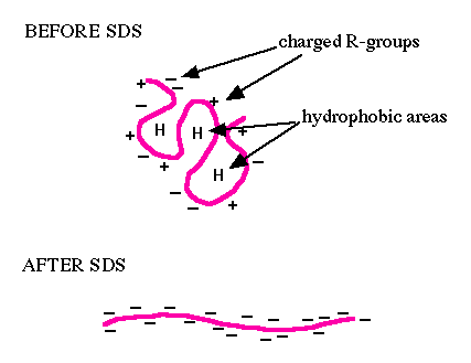
Figure 1. This cartoon depicts what happens to a protein (pink line) when it is incubated with the denaturing detergent SDS. The top portion of the figure shows a protein with negative and positive charges due to the charged R-groups in the protein. The large H's represent hydrophobic domains where nonpolar R-groups have collected in an attempt to get away from the polar water that surrounds the protein. The lower diagram shows that SDS can disrupt hydrophobic areas and coat proteins with many negative charges which overwhelms any positive charges the protein had due to positively charged R-groups. The resulting protein has been denatured by SDS (reduced to its primary structure) and as a result has been linearized.
PAGE
If the proteins are denatured and put into an electric field, they will all move towards the positive pole at the same rate, with no separation by size. So we need to put the proteins into an environment that will allow different sized proteins to move at different rates. The environment of choice is polyacrylamide, which is a polymer of acrylamide monomers. When this polymer is formed, it turns into a gel and we will use electricity to pull the proteins through the gel so the entire process is called polyacrylamide gel electrophoresis (PAGE). A polyacrylamide gel is not solid but is made of a laberynth of tunnels through a meshwork of fibers (figure 2 and figure 3).
If the proteins are denatured and put into an electric field, they will all move towards the positive pole at the same rate, with no separation by size. So we need to put the proteins into an environment that will allow different sized proteins to move at different rates. The environment of choice is polyacrylamide, which is a polymer of acrylamide monomers. When this polymer is formed, it turns into a gel and we will use electricity to pull the proteins through the gel so the entire process is called polyacrylamide gel electrophoresis (PAGE). A polyacrylamide gel is not solid but is made of a laberynth of tunnels through a meshwork of fibers (figure 2 and figure 3).
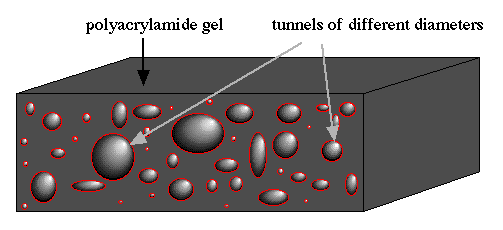
Figure 2. This cartoon shows a slab of polyacrylamide (dark gray) with tunnels (different sized red rings with shading to depict depth) exposed on the edge. Notice that there are many different sizes of tunnels scattered randomly throughout the gel.
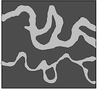
Figure 3. This is a top view of two selected tunnels (only two are shown for clarity of the diagram). These tunnels extend all the way through the gel, but they meander through the gel and do not go in straight lines. Notice the difference in diameter of the two tunnels.
Now we are ready to apply the mixture of denatured proteins to the gel and apply the current (figure 4). If all the proteins enter the gel at the same time and have the same force pulling them towards the other end, which ones will be able to move through the gel faster? Think of the gel as a tiny forrest with many branches and twigs througout the forest but they form tunnels of different sizes. If we let children and adults run through this forest at the same time, who will be able to get through faster? The children of course. Why? Because of their small size, they move through the forest faster since they have access to more of the paths in the forest while adults are limited to only the larger paths. Likewise, small molecules can manuver through the polyacrylamide forest faster than big molecules.
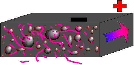
Figure 4. Cartoon showing a mixutre of denatured proteins (pink lines of differen lengths) beginning their journey through a polyacrylamide gel (gray slab with tunnels). An electric filed is established with the positive pole (red plus) at the far end and the negative pole (black minus) at the closer end. Since all the proteins have strong negative charges, they will all move in the direction the arrow is pointing (run to red).You have to remember that when we work with proteins, we work with many copies of each type of protein. As a result, the collection of proteins of any given size tend to move through the gel at the same rate, even if they do not take exactly the same tunnels to get through. Back to our analogy of the forest... If we were in a hot air ballon above the forest and watched 100 children, 100 teenagers, and 100 large adults running through the forest, we would see a collection (or band) of children moving quickly though each individual would choose his or her own route through the forrest. Behind the smallest individuals, we would see a band of teenagers moving slower, and a third band made of adults plodding their way through the forest using only the largest of paths. Likewise, proteins tend to move through a gel in bunches, or bands, since there are so many copies of each protein and they are all the same shape and size. When running an SDS-PAGE, we never let the proteins electrophorese (run) so long that they actually reach the other side of the gel. We turn off the current and then stain the proteins and see how far they moved through the gel (until we stain them, they are colorless and thus invisible). Figure 5 shows a cartoon gel and figre 6 shows a real one. Notice that the actual bands are equal in size, but the proteins within each band are of different sizes.
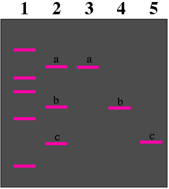
Figure 5. Top view of an SDS PAGE after the current has been on for a while (positive pole at the bottom) and then turned off. The gel (gray box) has five numbered lanes where five different samples of proteins (many copies of each kind of protein) were applied to the gel. (Lane 1, molecular weight standards of known sizes; Lane 2, a mixture of three proteins of different sizes with a being the largest and c being the smallest protein; Lane 3, protein a by itself; Lane 4, protein b by itself; Lane 5 protein c by itself.) Notice that each group of the three proteins migrated the same distance in the gel whether they were with other proteins (lane 2) or not (lanes 3-5). The molecular weight standards are used to measure the relative sizes of the unknow proteins (a, b, and c).
Monday, January 10, 2011
Faster Approach Hastens Creation of Powerful DNA Targeting Tool
A team of researchers has developed a less labor-intensive way to create synthetic enzymes that target specific DNA sequences for inactivation, repair, or alteration.
The study, performed by researchers from the molecular pathology unit of Massachusetts General Hospital (MGH; Boston, MA, USA), was published December 2010 online in the journal Nature Methods, and described a highly effective but less labor-intensive way to generate powerful tools called zinc-finger nucleases (ZFNs).
“With our approach, called context-dependent assembly, any scientist can use either standard molecular biology techniques or commercial DNA synthesis to design ZFNs for their target gene of interest,” said J. Keith Joung, MD, PhD, associate chief for research in MGH pathology, the study’s senior author. “ZFNs are broadly applicable, powerful tools for manipulating the genomes of cells from various organisms--including humans--and may provide a way to efficiently correct gene mutations responsible for human disease, avoiding problems resulting from the imprecise nature of current gene therapy approaches using viral vectors.”
Most human transcription factors that control whether a genetic signal is converted into a protein bind to specific DNA sequences using peptides called zinc fingers. Zinc-finger nucleases are synthetic “designer” proteins combining a zinc-finger domain, engineered to bind a specific DNA sequence, with an enzyme that breaks both DNA strands at the targeted site. While ZFNs have great potential, creating the customized proteins has been challenging.
In the simplest approach, called modular assembly, individual peptides are linked together similar to beads on a string to create a multifinger protein hypothetically able to recognize long DNA segments. Dr. Joung and others have shown that, in practice, modular assembly has a very low success rate for creating multifinger proteins. This high failure rate is most likely caused by “context-dependent” effects that individual zinc fingers can have on the DNA-binding activities of their neighboring fingers. Assembling peptides that do not work well together would be like trying to put together jigsaw puzzle pieces that do not fit.
In 2008, Dr. Joung and colleagues at the University of Minnesota (Twin Cities, USA) and other institutions, members of the Zinc Finger Consortium, reported developing a method called OPEN (Oligomerized Pool ENgineering), which takes these context-dependent effects into account. But although OPEN works well, it can be labor-intensive and very time consuming--requiring up to one year for a lab to establish the technology and two months of work to create desired ZFNs. To address these limitations, the MGH research team has assembled an extensive archive of zinc fingers known to work well when positioned together--basically, puzzle pieces that already have been put together. Using this context-dependent technique, the investigators were able to assemble dozens of ZFNs in as little as four days.
“With this archive in hand, any researcher can easily generate their own ZFNs in less than a week, and no special expertise is needed,” Dr. Joung explained. “In addition to being much faster, context-dependent assembly can generate large numbers of ZFNs simultaneously, which is hard to do with OPEN because it is more labor intensive.”
As was the case with OPEN, the Joung lab and the Zinc Finger Consortium will make the software and reagents required to practice context-dependent assembly available to all academic laboratories.
“One of the holy grails of genetics is the ability to make targeted changes to individual genes,” stated Laurie Tompkins, PhD, who over sees genetics grants at the National Institute of General Medical Sciences, one of the US National Institutes of Health (Bethesda, MD, USA) and a major supporter of this study. “Dr. Joung and his colleagues have developed an extraordinarily simple, efficient strategy for using zinc finger technology to swap out altered versions of genes for normal ones--or vice versa--providing basic scientists and clinicians alike with a broadly applicable research tool.”
Added Dr. Joung, an associate professor of pathology at Harvard Medical School (Boston, MA, USA), “At this point, I believe that context-dependent assembly will have the biggest impact on researchers using ZFNs to genetically manipulate model organisms, possibly even models developed from pluripotent stem cells. Other big impacts should be enabling researchers to create knockout mutations in a large series of genes involved in a common pathway or related to a specific disease and to use ZFNs to create comprehensive collections of mutants for every gene in an organism.”
Dr. Joung is also a member of the MGH Center for Computational and Integrative Biology and Center for Cancer Research. The challenges presented to scientists interested in using ZFNs in their investigations were described in an article in the Fall 2010 issue of the MGH-sponsored magazine Proto.
Sunday, January 9, 2011
Natural Plant Compound Fights Inflammation
Researchers have discovered how abscisic acid, a natural plant hormone with known beneficial properties for the treatment of disease, helps combat inflammation. The findings reveal significant new drug targets for the development of treatments for inflammatory and immune-mediated diseases.
The scientists, from Virginia Bioinformatics Institute at Virginia Tech (Blacksburg, USA), published their results in the November 2010 issue of the Journal of Biological Chemistry. They had reported some of the major molecular events in the immune system of mice that contribute to inflammation-related disease, including the involvement of a specific molecule found on the surface of immune cells involved in the body’s fight against infection. They have now gone a step further and revealed the process by which the natural drug abscisic acid interacts with this protein, known as peroxisome proliferator-activated receptor-gamma, to block inflammation and the consequent onset of disease.
"In previous work, our research group demonstrated that abscisic acid has beneficial effects on several conditions and diseases including obesity-related inflammation, diabetes, atherosclerosis, and inflammatory bowel disease,” said Dr. Josep Bassaganya-Riera, associate professor of immunology at the Virginia Bioinformatics Institute, leader of the Nutritional Immunology and Molecular Medicine Group in the institute’s cyberInfrastructure division, and lead investigator of the study. “One idea for how abscisic acid reduces inflammation in these instances is that it binds to a special region of peroxisome proliferator-activated receptor-gamma, a binding site known as the ligand-binding domain where the drug would be expected to latch on to and exert its effect. Our results show that this is not the case and, for the first time, we have demonstrated that abscisic acid works independently of this ligand-binding domain of the receptor.”
“The outcomes of this research illustrate the synergism that can result from combining computational and experimental approaches to characterize therapeutic targets,” said Dr. David Bevan, associate professor of biochemistry at Virginia Tech. “By using molecular modeling approaches we were able to identify a potential binding site for abscisic acid on the lanthionine synthetase C-like 2 protein, a protein required for the beneficial health effects of abscisic acid. We were also able, again using docking studies, to reveal reasons for the lack of direct association of abscisic acid with peroxisome proliferator-activated receptor-gamma, which was experimentally validated by ligand-binding assays.”
“This information is significant because it suggests the existence of new therapeutic targets or alternative modes of action that account for the effects of abscisic acid in the immune system,” added Dr. Bassaganya-Riera. “Drugs that bind to the ligand-binding domain of peroxisome proliferator-activated receptor-gamma such as Avandia are associated with severe cardiovascular side effects. In contrast, the newly discovered alternative mechanism of peroxisome proliferator-activated receptor-gamma activation by abscisic acid does not appear to be linked to any known adverse side effects, thereby representing a promising new therapeutic avenue."
“Lanthionine synthetase C-like 2 represents the first step in a pathway leading to activation of peroxisome proliferator-activated receptor-gamma in immune cells by abscisic acid,” said Dr. Raquel Hontecillas, assistant professor of immunology at the Virginia Bioinformatics Institute and one of the lead investigators of the study. “We have also shown that abscisic acid affects the expression of several genes involved in inflammation, metabolism and cell signaling, which provides further clues for possible intervention points in the treatment of inflammatory and immune-mediated diseases.”
The researchers plan to isolate more closely some of the new drug targets in the molecular network of the immune response as they continue to dissect the way that the naturally occurring drug abscisic acid reduces damage due to inflammation. Moreover, this new understanding on how abscisic acid works will be used to develop new classes of drugs that target the same alternative pathway of peroxisome proliferator-activated receptor-gamma activation, a potentially safer method than the use of drugs that target direct binding to the receptor.
Subscribe to:
Posts (Atom)